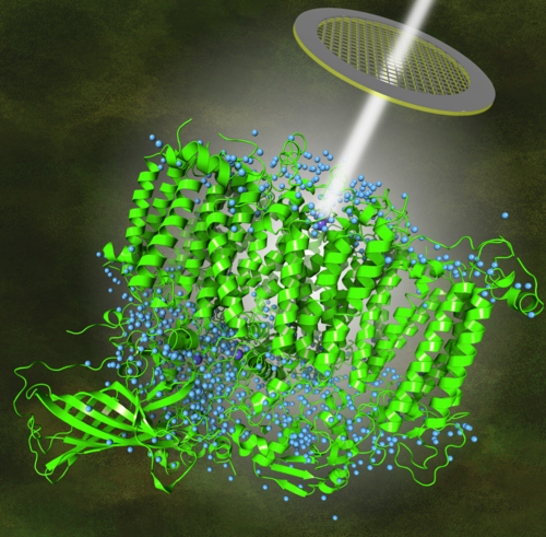
More than half of expected hydrogen atoms in PSII discovered by Hussein et al (project A05, published in Science)
Cryo-electron microscopy of PSII visualized water molecules and protons at 1.71 angstroms.
Image Credit: Dr. Dmitry Shevela (SciGrafik)
Project A05 is focussing their research on the protein complex photosystem II. In a new Science paper they used cryo-electron microscopy to visiualize PSII at even higher resolution than existing x-ray structures to identify protons. Hussein et al. collected high-resolution cryo–electron microscopy data of fully hydrated photosystem II from the thermophilic cyanobacterium Thermosynechococcus vestitus to a final resolution of 1.71 angstroms. The structure reveals several previously undetected partially occupied water binding sites and more than half of the hydrogen and proton positions. This clarifies the pathways of substrate water binding and plastoquinone B protonation.
News from Jun 20, 2024
Cryo-electron microscopy reveals hydrogen positions and water networks in photosystem II
The advancements of cryo-EM have allowed a transformative change in the field of structural biology, enabling the capture of atomic resolution three-dimensional structural data from highly purified and homogeneous protein complexes flash-frozen from their dispersions in aqueous buffer (23). Although concerns presently remain regarding radiation damage to high-valent metal centers, these should not distract from the exceptional insights provided by this technique.
The present 1.71 Å–resolution cryo-EM structure of dimeric PSII from T. vestitus unveils partially occupied water-binding sites near the water-splitting Mn4CaO5 cluster, strongly supporting water delivery to the Mn4CaO5 cluster through the O1 channel. Similarly, it provides experimental detection of W63 in intact PSII. By confirming this crucial structural feature of a theoretical calculation (22, 35, 38), our data settles the protonation pathways of QBH– and for the subsequent reprotonation of the proton donor (Fig. 5E).
A notable feature of cryo-EM is its enhanced scattering potential for light elements, such as hydrogen, which offers distinct advantages over x-ray crystallography at similar resolution. Our analysis shows >50% of the hydrogen or proton positions within PSII, which greatly extends present structural models. Knowledge of the protonation states of amino acids, cofactors, and/or substrates near the catalytic sites of PSII is essential for revealing how their complex interactions enable efficient proton-coupled electron transfer and water oxidation. The various degrees of proton or hydrogen detection in different regions of the protein are suggestive of local protein dynamics that may play a role in catalysis, exemplified by the marked differences in the QA and QB binding sites.
Publication
Hussein, R., Graca, A., Forsman, J., Aydin, A.O., Hall, M., Gaetcke, J., Chernev, P., Wendler, P., Dobbek, H., Messinger, J., Zouni, A. and Schröder, W.P. (2024). Cryo-electron microscopy reveals hydrogen positions and water networks in photosystem II. Science, 384, 6702: 1349-1355. doi: 10.1126/science.adn6541.
